Viruses under a microscope. What viruses look like Photos of unusually beautiful viruses
Medical luminaries are working to create a cure against the human immunodeficiency virus. To understand the nature of the disease and the characteristics of its spread, scientists need to know what a virus cell looks like.
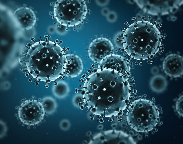
The structure of the virus is like a sphere that is covered with spikes. Its size significantly exceeds the parameters of the causative agent of hepatitis B and other viruses. The diameter of the sphere is 100 - 150 nanometers. It is called the nucleocapsid, or virion.
The cellular structure of HIV is characterized by a two-layer structure:
- shell covered with “spikes”;
- the cell body that contains nucleic acid.
Together they make up a virion - a particle of the virus. Each of the “spikes” covering the shell looks like a mushroom with a thin stem and cap. With the help of these “mushrooms,” the virion interacts with foreign cells. Surface glycoproteins (gp120) lie on the surface of the caps. Other glycoproteins, transmembrane (gp41), are located inside the “legs”.
At the heart of the viral cell lies a genome - RNA, consisting of 2 molecules. Each of them stores 9 genes that carry information about the structure of the virus, methods of infection and reproduction of harmful cells.
The genome is surrounded by a conical shell, which consists of proteins:
- p17- matrix;
- p24 - capsid.
The genomic RNA is associated with the envelope through the nucleocapsid proteins p7 and p9.
There are several known forms of the human immunodeficiency virus. The most common of them is HIV-1. It is distributed in Eurasia, North and South America. Another form of HIV-2 has been identified in the population of the African continent. HIV-3 and HIV-4 are rare.
What family does the HIV virus belong to?
HIV belongs to the family of retroviruses - their virions contain RNA that attacks the body of vertebrates. Once in the body, virions cause the death of healthy cells. Retroviruses infect animals. Only one species in this family is dangerous to humans -.
This virus belongs to the group of lentiviruses. Translated from Latin, “lentus” means “slow.” From the name it is clear that diseases caused by these microorganisms have a long course and a long incubation period. After HIV DNA has entered the human body, it may take 5-10 years until the first signs of the disease appear.
Since the mid-80s of the 20th century, studies studying the HIV genome have appeared in genetics. Scientists have not yet found a way to completely destroy HIV cells, but have made great strides in diagnosing and treating the disease. The use of antiretroviral drugs can extend the latent stage of the disease to 15 years. The life expectancy of patients is constantly increasing. Today it averages 63 years.
What HIV looks like under a microscope
Pictures of a manifold enlarged HIV were first taken in 1983. The elementary unit of HIV under a microscope resembles a model of a mysterious planet, which is covered with exotic plants. Thanks to the development of photographic and optical equipment, detailed photographs of the dangerous viral particle were later taken.
Computer graphics allow you to reproduce its life cycle:
- At the stage of release of the virion from the cell, the image shows convex seals that seem to be bursting the cell from the inside.
- At first, after separation, the virus has a process that connects it to the cell. It gradually disappears.
- When the stage of virus isolation from the cell is completed, it takes the shape of a ball. Appears as a black ring in a macro photograph.
- A mature virion in the photo looks like a black rectangle, triangle or circle that is framed by a thin ring. The dark core is the capsid. It has the shape of a cone. Which geometric figure will be visible in the photograph, depends on the angle from which the picture was taken. The ring is the shell of the virion.
Which cells and in what quantity are affected?
The cellular receptors that the viral protein binds to are called CD4. Elementary units of a living organism that have such receptors are potential targets for HIV. The CD4 protein receptor is part of some leukocytes, namely T-lymphocytes, monocytes and macrophages.
T-lymphocytes (helpers), protecting the body, are the first to come into contact with aggressive virions and die. In a healthy person, CD4 is detected in the amount of 5–12 units per blood sample. With the development of infection, the norm drops to 0 - 3.5 units.
After the immunodeficiency virus penetrates the internal environment of the body, changes in cells do not occur immediately. It takes time for dangerous viruses to grow stronger and adapt to the environment. This takes at least a week. Next, the viral particle, with the help of “fungi” covering its surface (gp160), clings to the CD4 receptors of healthy cells. Then they invade under the membrane shell.
Being under the shell of lymphocytes, macrophages, nerve cells, the invader viruses hide from the effects of drugs and the resistance of the immune system. They disrupt the immune responses of the body, which begins to react to its own cells as foreign antigens.
Inside the affected cells, the immunodeficiency virus multiplies with the subsequent release of new virions. The host cell is destroyed.
When cells are attacked by an immunodeficiency virus, a protective reaction is triggered. Gradually, the immune system forms antibodies to the virus. Their number increases and after 2 - 3 weeks the antibodies will be noticeable in an enzyme immunoassay of blood. If a small number of viral particles have entered the body, a sufficient number of antibodies can be formed only after a year. This happens in 0.5% of cases.
Thus, information about the structure and activity of the immunodeficiency virus helps scientists in the development of diagnostic methods and methods of treating HIV infection.
Viruses are too small to be seen under a regular microscope. Therefore, they are examined under an electronic microscope.
Let's take a look at some of them:
Here we see two viruses - rotavirus is shown with an arrow on the left, adenovirus is shown on the right. It can be seen that the adenovirus has the shape of an icosahedron, and the rotavirus is a ball covered with pimples. Both are found in the stool of a child with diarrhea (caused by rotavirus). 
By the way, notice technological progress. An adenovirus is also depicted here, but the details are almost impossible to discern. This article is from 1984, and the one above was from 2003.

Parvovirus. It is significantly smaller than the previous two (the scale is shown in both pictures). Even under electron microscope parvovirus is difficult to see.

And this is a relative of smallpox. Oval in shape, approximately 200 nm in length.

Polio virus. Very small, but very malicious. RNA enclosed in a protein shell. Scale: 100 nm.

Influenza virus. Covered with a lipid shell, ordered internal structure does not have. It can be seen that its entire surface is covered with shell proteins - HA and NA. Scale: 100 nm.

And these are cells infected with the Nodamura virus. The virus assembles and accumulates inside cells; it does not need to be covered by a cell membrane. When the cell becomes overcrowded, it bursts and the viruses come out.

And these are cells infected with the SARS virus. It assembles its capsid near the cell membrane and then buds off from the cell, carrying with it a piece of its membrane. Therefore new viral particles gradually emerge from the cell. The arrow shows the viral particle in the process of assembly. It can be seen that viral particles are in the space between the cells.

Quite old, but not bad photo
Last week, the Russian company Visual Science showed the world's first atomic resolution model of the Zika virus. According to the creators, the images made on the basis of this model are the most detailed and scientifically reliable of all that exist today. The company has been working on viruses for several years now; its “Zoo” includes HIV, Ebola, influenza, and many others. We talked with the founder of the company, Ivan Konstantinov, about the standards by which Visual Science works and why the authors call their models the most accurate in the world.
When did work on creating a model of the virus begin and how long did it take?
This is a record-breaking job for us. Large, complex viruses typically take eight to nine months of simulation to complete. In the case of Zika, we threw all our resources into the job and completed it in three weeks.
I understand that interest in this virus is related to the current outbreak and possible epidemic of the virus in North America?
In general, we have never made our models for any global events, “on occasion”. Even the Ebola virus, the image of which was used by almost all the world's media during the recent outbreak in 2014, was made long before the outbreak itself. We choose viruses based on how common and dangerous they are, and how interesting their structure is. And, of course, how well it has been studied. But the Zika virus model is our first experience of such “high-speed” modeling.
How many people worked on the model?
(counts) Eight people.
You talk about your models as “the most scientifically accurate images” that exist today. This raises two questions. Firstly, what does this mean - what do you mean by the concept of “scientific validity” in relation to the model? And, secondly, how is this reliability achieved?
First of all, it should be said that our company was founded by molecular biologists and bioinformatics specialists. Therefore, our scientific background determines how we approach modeling. Our modeling is based on an emphasis on scientific expertise, which allows us to avoid the mistakes that abound in models if they are worked exclusively by designers and artists. Where there is no scientific component in working on a model.
When creating each virus, we work according to the following scheme. First, we analyze all available publications that relate to its structure, on the basis of which a global literature review is compiled, formed according to certain internal standards. We determine which of the components of the virus have been fully studied - which of them have X-ray data - and which of them have not been studied. For each component, we determine which of its fragments are not in the X-ray structures. The X-ray structural analysis method suggests that many regions of the protein that are difficult to crystallize and complicate the production of crystals - transmembrane fragments, regions with a mobile structure, and so on - can be specially cut off by scientists, so that they are simply not in the published data. We find these missing fragments and complete their structure based on the methods that are currently used for this in science (this is data on the structure of related proteins and molecular dynamics methods).
At the next stage, we predict protein-protein interactions inside the virus: which proteins and which surfaces contact each other in the virion, how exactly the protein complexes are structured. X-ray structural analysis usually does not provide information about this, so we turn to molecular docking methods.
It happens when the structure of the components of a certain virus is not yet known to us at all. In this case, it is necessary to use data on related viruses as templates for a new structure. This is also a fairly common technique in science; special protocols have been developed for this, which we use in modeling.
The most controversial aspect of the model is almost always the packaging of the virus genome. This is a very difficult problem, which often simply cannot be solved unambiguously by existing methods. That's why we always say that we only show in the model possible variant genome folding. We, of course, try to predict DNA-protein or RNA-protein interactions for the genome as completely as possible, and try to establish using bioinformatics methods the elements of the tertiary spatial structure of RNA genomes. But it is simply impossible to do this absolutely reliably now. This is an independent, large and interesting scientific problem.
Plus, for each project we select the most authoritative experts who have devoted many years to studying a particular virus, and we consult with them. In the case of the influenza virus, for example, it was the group of Jaime Martin-Benito from the Spanish National Center of Biotechnology and a number of other researchers. For HIV it was Egor Voronin from Global HIV Vaccine Entreprise. When modeling Ebola, we talked with Ronald Harty from the University of Pennsylvania.
What about Zika?
The Zika virus is a rather exotic thing; it has long been deprived of the attention of scientists. In this case, there were no large scientific groups that would be leaders in this matter and could help us. Fortunately, this virus is quite simple, and in this case there was no particular need to involve third-party consultants; we managed it on our own. After the model was ready, we received a number of minor comments regarding, for example, the depth of immersion of transmembrane protein fragments into the membrane.
What was the most difficult thing when creating this model?
Probably the spatial organization of genome packaging. It's always difficult.
In the case of the Zika virus, what percentage of proteins already have X-ray structures, and what did you have to reconstruct using analogical modeling methods?
So far, crystallographic data on the protein structures of the Zika virus itself are not available, so all spatial models were made based on genome reads and related proteins from the related Dengue, West Nile, or yellow fever viruses.
The Zika virus is the latest addition to your Virus Zoo. Please tell us what kind of project this is and what goals you set for it.
Zoo is the world's first attempt to create a collection of scientifically accurate models of human viruses at atomic resolution. The fact is that viruses are too small to study them using methods suitable for studying cells. On the other hand, they are too large to work with as proteins and obtain X-ray structures. It turns out that we know quite a lot about the structure of individual viral components and how viruses behave in cells. But you can see viruses with your own eyes and examine them in all details only with the help of computer modeling.
There is even some paradox here: any child knows what Mars, far from us, looks like, but few know what the flu virus, which we all get sick with every year, looks like. Actually, from this desire - to show people the beauty and complexity of the micro and nanoworld of the viral world - our “Zoo of Viruses” grew. This is, first of all, a purely educational and educational project, and we are glad that many of our images have already appeared on the pages of leading textbooks and guides.
I can’t help but ask: will my favorite giant mimivirus end up in “Zoo”?
Mimivirus is, of course, very large. True, it’s big, it doesn’t infect humans, and “Zoo of Viruses” is primarily about human viruses. And, besides, it is rather poorly studied. We must understand that our models represent all the molecules that are in the virion, even individual membrane lipids. Therefore, even in the case of Ebola, the number of molecules is already in the millions, and modeling requires very large resources. In this sense, the mimivirus, which is smaller in size than some bacterial cells, looks, of course, terrifying. Apparently, to model it we will need to develop a special approach, so this is not a task for the near future. For now, we are focused on more studied and widespread viruses - the herpes virus, hepatitis and a dozen others.

Ebola virus

Influenza A/H1N1 Virus

AIDS virus

Human papillomavirus - HPV

Pathogenic microorganisms (viruses, bacteria and many others) can cause severe infectious diseases by overcoming the natural resistance of the human body.
Infectious diseases are caused by living organisms that can change and evolve. This process occurs much faster in microorganisms than in humans, allowing viruses and bacteria to find new ways to resist drugs.
tuberculosis bacterium, magnified 10 thousand times. Tuberculosis is a widespread infectious disease of humans and animals in the world, caused by various types mycobacteria, usually species Mycobacterium tuberculosis complex.

Microscopic particles of Coronaviruses. Coronaviruses is a family of about eleven types of viruses that infect humans, cats, birds, dogs, cattle and pigs.

Neisseria meningitidis bacterium, which causes a serious disease - meningococcal meningitis. In the picture, the bacterium is magnified 33 thousand times.

Anthrax bacterium (Bacillus) stem, magnified 18 thousand 300 times. Anthrax (malignant carbuncle, anthrax) is a particularly dangerous infectious disease of agricultural and wild animals of all types, as well as humans.

This colorful picture is actually - Ebola virus. Causes Ebola hemorrhagic fever. It multiplies so quickly that the affected cells of the body turn into crystal-like blocks of compacted virus particles.

Influenza virus, consisting of ribonucleic acid surrounded by a nucleocapsid (red) and a lipid envelope (green). The image is enlarged 230 thousand times. Influenza A viruses infect humans and some species of animals (horses, pigs) and birds. Influenza viruses types B and C are pathogenic only for humans.

Smallpox- one of the oldest diseases. In the past, it was the most common and most dangerous disease.

Smallpox viruses- the largest viruses containing DNA, the molecular weight of which is greater than that of any other animal virus.

Color image papillomavirus, which is the cause of warts in humans. The image is enlarged 60 thousand times.

Polio Virus: The RNA genetic material originates in the core of each virus, surrounded by a protein coat (blue). Poliomyelitis is infantile spinal paralysis, an acute infectious disease caused by damage to the gray matter of the spinal cord by poliovirus.

Colored, scanned micrograph of a bacterium spirochetes Borrelia Burgdorferi, which can cause Lyme disease in a person injured by a tick bite. Lyme disease is a disease that primarily affects the skin, nervous and cardiovascular systems, and the musculoskeletal system, and is prone to long-term progression.


Escherichia coli bacterium, which under certain conditions can cause gastroenteritis and urinary tract infections. Escherichia coli is a rod-shaped bacterium that belongs to the group of facultative anaerobes (it lives and reproduces only in the absence of direct oxygen). E. coli has many strains, most of which belong to the natural microflora of the human intestine and help prevent the development of harmful microorganisms and synthesize vitamin K. But some of its varieties can cause serious poisoning, intestinal dysbiosis and colibacillosis.

Pneumococcus bacterium, which can cause pneumonia of the upper respiratory tract in a person with immunodeficiency. Pneumococcus is the leader among all pathogens of certain respiratory diseases.

HIV AIDS) under a microscope. HIV is the human immunodeficiency virus that causes HIV infection, a disease whose final stage is known as acquired immunodeficiency syndrome (AIDS).
There is an opinion that animals, plants and humans predominate in numbers on planet Earth. But this is actually not the case. There are countless microorganisms (microbes) in the world. And viruses are among the most dangerous. They can cause various diseases in humans and animals. Below is a list of the ten most dangerous biological viruses for humans.
Hantaviruses are a genus of viruses that are transmitted to humans through contact with rodents or their waste products. Hantaviruses cause various diseases belonging to such groups of diseases as “hemorrhagic fever with renal syndrome” (mortality on average 12%) and “hantavirus cardiopulmonary syndrome” (mortality up to 36%). The first major outbreak of disease caused by hantaviruses, known as Korean hemorrhagic fever, occurred during the Korean War (1950–1953). Then more than 3,000 American and Korean soldiers felt the effects of a then unknown virus that caused internal bleeding and impaired kidney function. Interestingly, this particular virus is considered probable cause the outbreak of an epidemic in the 16th century that exterminated the Aztec people.
Influenza virus is a virus that causes an acute infectious disease of the respiratory tract in humans. Currently, there are more than 2 thousand of its variants, classified into three serotypes A, B, C. The group of viruses from serotype A, divided into strains (H1N1, H2N2, H3N2, etc.) is the most dangerous for humans and can lead to epidemics and pandemics. Every year, between 250 and 500 thousand people worldwide die from seasonal influenza epidemics (most of them children under 2 years of age and elderly people over 65 years of age).

Marburg virus - dangerous virus first described in 1967 during small outbreaks in the German cities of Marburg and Frankfurt. In humans, it causes Marburg hemorrhagic fever (mortality rate 23-50%), which is transmitted through blood, feces, saliva and vomit. The natural reservoir for this virus is sick people, probably rodents and some species of monkeys. Symptoms in the early stages include fever, headache and muscle pain. In the later stages - jaundice, pancreatitis, weight loss, delirium and neuropsychiatric symptoms, bleeding, hypovolemic shock and multiple organ failure, most often the liver. Marburg fever is one of the top ten deadly diseases transmitted from animals.

Sixth on the list of the most dangerous human viruses is Rotavirus, a group of viruses that are the most common cause of acute diarrhea in infants and young children. Transmitted by the fecal-oral route. The disease is usually easy to treat, but kills more than 450,000 children under five worldwide each year, most of whom live in underdeveloped countries.
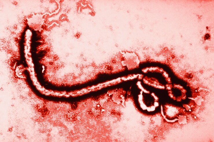
Ebola virus is a genus of virus that causes Ebola hemorrhagic fever. It was first discovered in 1976 during an outbreak of the disease in the Ebola River basin (hence the name of the virus) in Zaire, DR Congo. It is transmitted through direct contact with the blood, secretions, other fluids and organs of an infected person. Ebola fever is characterized by a sudden increase in body temperature, severe general weakness, muscle pain, headaches, and sore throat. Often accompanied by vomiting, diarrhea, rash, impaired renal and liver function, and in some cases internal and external bleeding. According to the US Centers for Disease Control, in 2015, 30,939 people were infected with Ebola, of whom 12,910 (42%) died.

Dengue virus is one of the most dangerous biological viruses for humans, causing dengue fever, in severe cases, which has a mortality rate of about 50%. The disease is characterized by fever, intoxication, myalgia, arthralgia, rash and swollen lymph nodes. It is found mainly in the countries of South and Southeast Asia, Africa, Oceania and the Caribbean, where about 50 million people are infected annually. The carriers of the virus are sick people, monkeys, mosquitoes and bats.

Smallpox virus is a complex virus, the causative agent of a highly contagious disease of the same name that affects only humans. This is one of the oldest diseases, the symptoms of which are chills, pain in the sacrum and lower back, rapid increase in body temperature, dizziness, headache, vomiting. On the second day, a rash appears, which eventually turns into purulent blisters. In the 20th century, this virus claimed the lives of 300–500 million people. About US$298 million was spent on the smallpox campaign from 1967 to 1979 (equivalent to US$1.2 billion in 2010). Fortunately, the last known case of infection was reported on October 26, 1977 in the Somali city of Marka.

The rabies virus is a dangerous virus that causes rabies in humans and warm-blooded animals, which causes specific damage to the central nervous system. This disease is transmitted through saliva from the bite of an infected animal. Accompanied by an increase in temperature to 37.2–37.3, poor sleep, patients become aggressive, violent, hallucinations, delirium, a feeling of fear appear, soon paralysis of the eye muscles, lower extremities, paralytic respiratory disorders and death occurs. The first signs of the disease appear late, when destructive processes have already occurred in the brain (swelling, hemorrhage, degradation of nerve cells), which makes treatment almost impossible. To date, only three cases of human recovery without vaccination have been recorded; all others ended in death.
Lassa virus is a deadly virus that is the causative agent of Lassa fever in humans and primates. The disease was first discovered in 1969 in the Nigerian city of Lassa. It is characterized by a severe course, damage to the respiratory system, kidneys, central nervous system, myocarditis and hemorrhagic syndrome. It is found mainly in West African countries, especially in Sierra Leone, the Republic of Guinea, Nigeria and Liberia, where the annual incidence ranges from 300,000 to 500,000 cases, of which 5 thousand lead to the death of the patient. The natural reservoir of Lassa fever is polymammated rats.

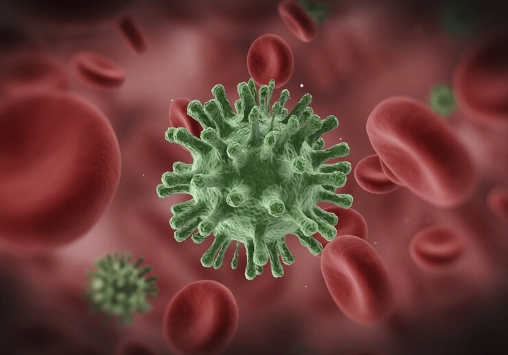
Human immunodeficiency virus (HIV) is the most dangerous human virus, the causative agent of HIV infection/AIDS, which is transmitted through direct contact of mucous membranes or blood with bodily fluid of the patient. During HIV infection, the same person develops new strains (varieties) of the virus, which are mutants, completely different in reproduction speed, capable of initiating and killing certain types of cells. Without medical intervention, the average life expectancy of a person infected with the immunodeficiency virus is 9–11 years. According to 2011 data, 60 million people worldwide have become infected with HIV, of which 25 million have died, and 35 million continue to live with the virus.